
In a study published online in Cell, a research group led by Prof. ZHANG Hong from the Institute of Biophysics of the Chinese Academy of Sciences demonstrated that Ca2+ transients on the endoplasmic reticulum (ER) surface trigger liquid-liquid phase separation (LLPS) of FIP200 to specify autophagosome initiation sites, which answers the long-standing question in autophagy—the nature of the signal that triggers the targeting of the autophagosome initiation complex to the ER.
Autophagy involves sequestration of cytoplasmic contents in autophagosomes and delivery to lysosomes for degradation and recycling. The molecular understanding of autophagy mainly originates from studying a set of autophagy-related (Atg) genes identified in yeast. Very little is known about the mechanisms underlying the unique steps in the much more complex autophagy pathway in multicellular organisms.
One of the fundamental differences between autophagy in yeast and multicellular organisms is that autophagosome initiation occurs at spatially distinct sites. In yeast, autophagosomes are generated on the vacuole membrane, while in multicellular organisms, autophagosomes are initiated simultaneously at multiple sites on the ER. The mechanism which initiates autophagosome formation on the ER in multicellular organisms is elusive.
The researchers reported that the Ca2+ transient on the ER cytosolic surface is essential for autophagosome initiation. They first developed a CYB5 ER transmembrane domain-tethered GCaMP6f biosensor with GCaMP6f facing the cytosolic side and found that GCaMP6f-CYB5 can be used as a Ca2+ biosensor to detect Ca2+ changes in a narrow spatial domain on the ER outer surface. They then performed multi-modal SIM (multi-SIM) analysis, which enables them to detect very rapid and localized changes in the GCaMP6f-CYB5 fluorescence signal, to examine the Ca2+ dynamics on the ER. Multi-SIM analysis detected that in starved or Torin1-treated cells, various forms of dynamic GCaMP6f-CYB5 signals in various regions of the cells were observed. These results indicated that autophagy induction triggers Ca2+ transients on the ER cytosolic surface.
Besides, the researchers identified that both attenuated and persistent Ca2+ oscillations on the ER outer surface cause a defect in autophagosome formation. In IP3R2/IP3R3 (inositol-1,4,5-triphosphate receptor) double-knockdown cells treated with Ca2+ release channel inhibitors (RCI), Ca2+ transients on the ER outer surface were greatly attenuated upon starvation. The formation of autophagosome initiating FIP200 puncta, and puncta formed by markers for autophagic structures such as WIPI2 (WD-repeat protein interacting with phosphoinositides) and LC3 was also greatly reduced, indicating that Ca2+ transients on the ER are essential for autophagosome initiation. They also found that release channel activator (RCA) treatment increased the frequency, amplitude and duration of local/global Ca2+ transients and oscillations on the ER, and caused accumulation of small-sized unacidified autophagic structures.
EPG-4/EI24 (ectopic PGL granules-4/etoposide-induced gene 2.4 kb) encodes a metazoan-specific ER transmembrane autophagy protein. Multi-SIM analysis revealed that the frequency and amplitude of ER Ca2+ transients were increased in EI24 KO cells. Puncta formed by FIP200, WIPI2 and LC3 were increased in EI24 KO cells, and this increase was suppressed by BAPTA-AM treatment. However, these autophagic structures are small-sized, unclosed autophagic structures. Thus, depletion of EI24 causes persistent Ca2+ oscillations on the ER outer surface and a characteristic autophagy defect, as in RCA treated cells.
In addition, the researchers used the multi-SIM system to examine the dynamic assembly of the autophagosome initiation complex in response to ER Ca2+ transients at a super-fast and super-resolution level in living cells. They found that Ca2+ transients on the ER surface induced formation of liquid-like FIP200 punctum which are highly dynamic and prone to fusion. FIP200 punctum attached to ER, moved along the ER tubules and coalesced with other FIP200 punctum until they reached up to 200~300 nm. The ER membrane proteins VAPs (VAMP-associated proteins A/B) and ATLs (Atlastin 2/3) stabilized the association of the FIP200 punctum complex with the ER. The FIP200 puncta on the ER then assembled into autophagosome initiation sites.
This study reveals a crucial role of Ca2+ transients at the ER/cytosol interface in triggering LLPS of FIP200 for specification of autophagosome initiation sites in multicellular organisms.
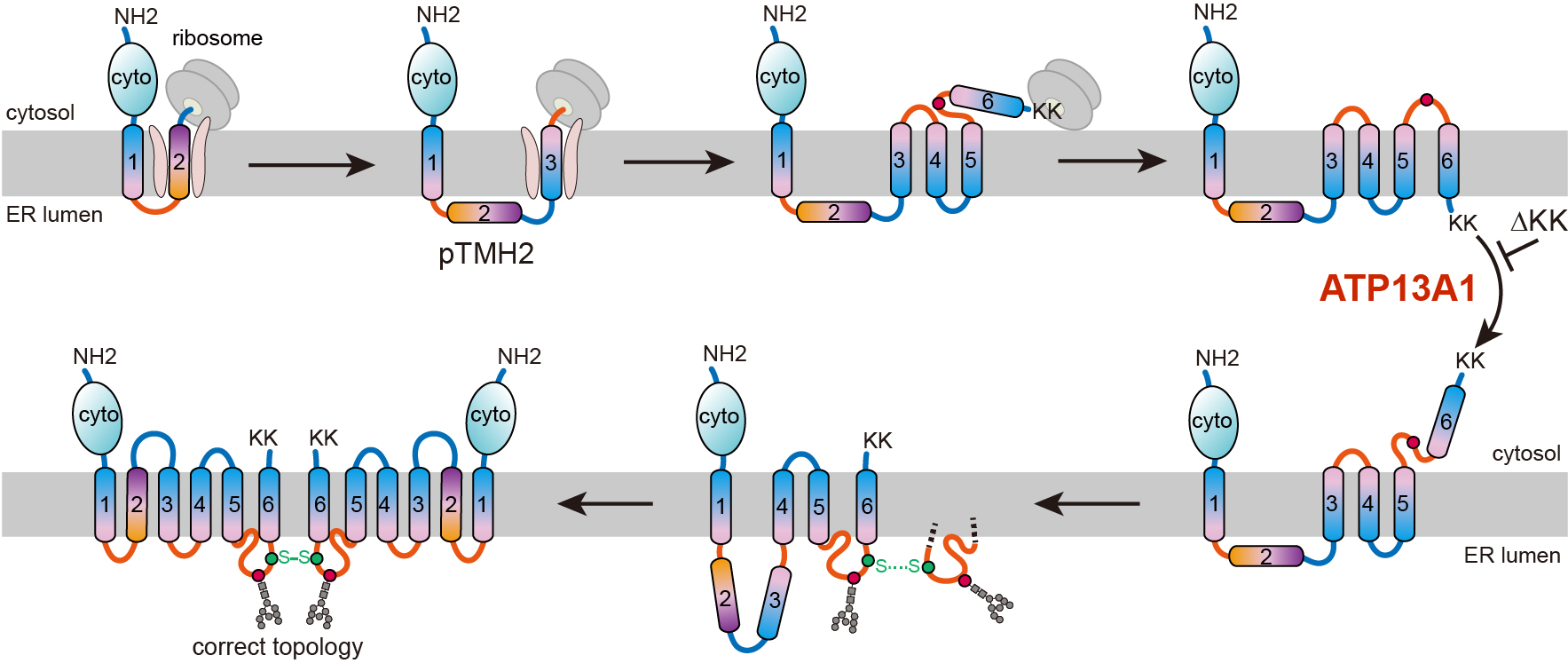
Researchers led by Prof. ZHANG Zairong from the Shanghai Institute of Organic Chemistry have identified a post-translational topogenesis pathway for the folding and assembly of multi-spanning membrane proteins.
Endoplasmic Reticulum-Associated Degradation (ERAD) is a special ubiquitin proteasome system located on endoplasmic reticulum (ER) and is responsible for removing misfolded proteins retained in the ER. Researchers from Dr. XIE Qi's group at the Institute of Genetics and D...

86-10-68597521 (day)
86-10-68597289 (night)

52 Sanlihe Rd., Xicheng District,
Beijing, China (100864)
Copyright © 2002 - Chinese Academy of Sciences


