
A research team led by Prof. CHEN Yan and Prof. YANG Lifeng from the Shanghai Institute of Nutrition and Health of the Chinese Academy of Sciences has revealed a physiological function of monocarboxylate transporter 1 (MCT1), which is coded by Slc16a1, in mediating intracellular and extracellular lactate transport in skeletal muscle. The team found that MCT1 regulates mitochondrial biogenesis of skeletal muscle and exercise activity. Furthermore, the deletion of Slc16a1 promotes the production of oxidative muscle fibers, enhances exercise endurance, and improves metabolic phenotypes. This study was published in Science Advances.
In the 1980s, Prof. George Brooks proposed the “lactate shuttle” theory, which posits that lactate serves as an energy substrate, produced by cells or tissues primarily relying on glycolysis for energy, but consumed by cells or tissues primarily utilizing oxidative metabolism. As the body’s largest organ for energy metabolism, skeletal muscle is both a major producer and consumer of lactate. The transport of lactate in skeletal muscle depends on the MCT family of proteins, especially MCT1 and MCT4. As early as 1998, scientists discovered that the expression of MCT1 in skeletal muscle is positively correlated with its oxidative capacity, while MCT4 shows a negative correlation.
Mammalian skeletal muscle mainly consists of four different types of muscle fibers, which are composed of different myosin types and metabolic characteristics. These muscle fibers include oxidative slow-twitch type 1 fibers, oxidative fast-twitch type 2A fibers, and glycolytic type 2X and 2B fibers. The metabolic characteristics of skeletal muscle depend on the proportion of these four types of muscle fibers. The higher the proportion of oxidative muscle fibers, the stronger the oxidative metabolic capacity of the skeletal muscle, and vice versa. However, before this study, the transport of lactate between different types of muscle fibers, as well as the specific distribution and function of MCT1 in different types of skeletal muscle fibers, had not yet been clearly elucidated.
In this study, researchers observed the specific distribution of monocarboxylate transporters MCT1 and MCT4 in skeletal muscle fibers, suggesting the possible existence of a “lactate shuttle” between muscle fibers dependent on MCT1 and MCT4.
Given the higher expression of MCT1 compared to MCT4 in skeletal muscle, researchers constructed an animal model with muscle-specific knockout of Slc16a1 (mKO) to investigate MCT1-mediated lactate metabolism in muscle fibers. They found that mKO mice exhibited an improvement in exercise endurance, an increase in the proportion of oxidative muscle fibers, and a decrease in the proportion of glycolytic muscle fibers. At the metabolic level, they also found that mKO mice had improved glucose tolerance, an increased metabolic rate, and enhanced utilization of glucose in the tricarboxylic acid (TCA) cycle in skeletal muscle, based on metabolic flux analysis and metabolomics combined with animal experiments.
Mechanistically, the researchers proposed that lactate transport in skeletal muscle fibers depended on two shuttles—a “cell-cell lactate shuttle” between muscle fibers and an “intracellular lactate shuttle” within oxidative muscle fibers. This hypothesis was validated through analyses with the mKO mice.
The researchers also found that normal lactate transport between muscle fibers depended on the combined involvement of MCT1 and MCT4. Lactate produced in glycolytic muscle fibers was transported extracellularly via MCT4, then taken up by oxidative muscle fibers through MCT1. Within oxidative muscle fibers, mitochondria also took up lactate via MCT1. Mitochondrial lactate dehydrogenase then converted lactate to pyruvate, consuming equivalent amounts of NAD+ and allowing pyruvate to enter the TCA cycle. In the absence of MCT1 in skeletal muscle, both lactate uptake by oxidative muscle fibers and its entry into mitochondria were impaired, leading to increased intracellular NAD+ levels. This increased the activity of SIRT1, an NAD+-dependent deacetylase, thus enhancing deacetylation and the activity of PGC-1α. Subsequently, mitochondrial biogenesis and function increased along with the formation of oxidative muscle fibers. As a result, phenotypes of muscle had a series of changes.
This study reveals the physiological roles of lactate transport at the muscle fiber and cellular levels. It not only validates and refines Prof. George Brooks’ “lactate shuttle” theory but also sheds light on the physiological processes of skeletal muscle, providing a new theoretical basis for the study of skeletal muscle physiology and pathology. Moreover, the conversion of skeletal muscle fiber types has long been a hot topic in exercise physiology research. Given the different metabolic characteristics of muscle fiber types and their varying resistance to damage and aging, as well as their contributions to different forms of exercise, this study offers a new direction for research in metabolic improvements, pathological damage, and exercise physiology.
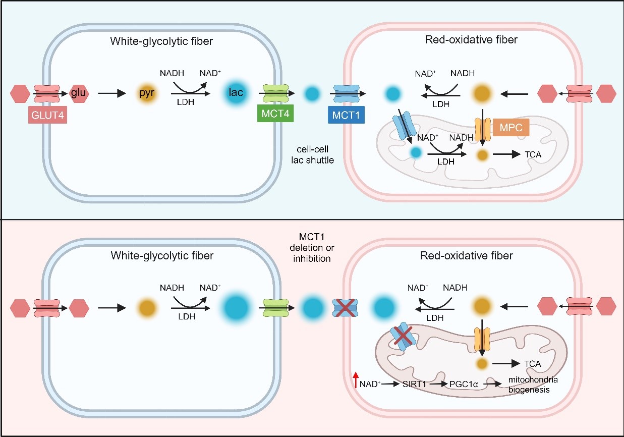
A model diagram of MCT1-mediated lactate shuttle in skeletal muscle. (Image by Prof. CHEN Yan’s group)

86-10-68597521 (day)
86-10-68597289 (night)

52 Sanlihe Rd., Xicheng District,
Beijing, China (100864)
Copyright © 2002 - Chinese Academy of Sciences


