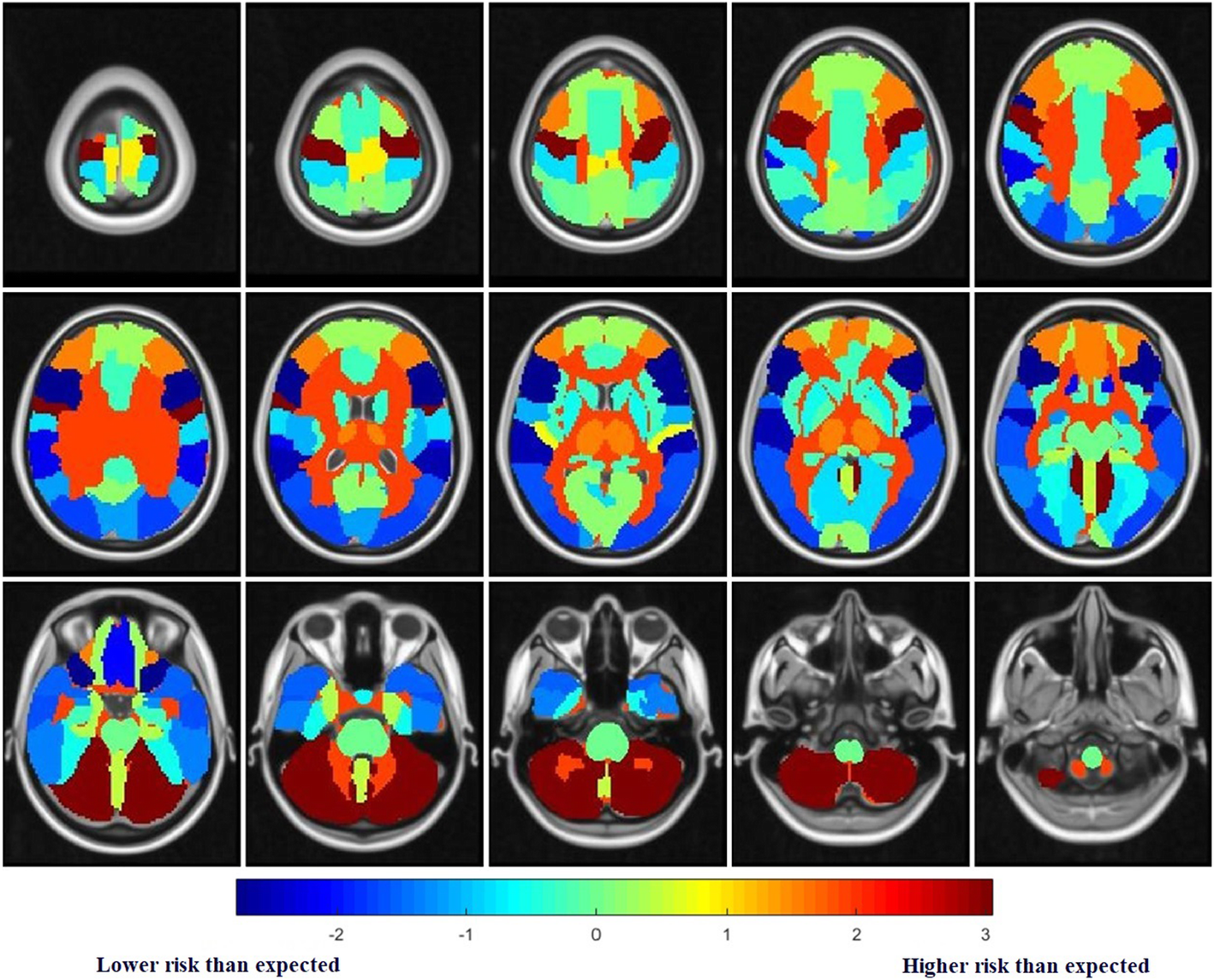
Recently, a brain metastasis risk map of small cell lung cancer (SCLC) has been developed based on the analysis of medical big data, which comprehensively showed the brain metastasis risk of SCLC in various brain regions. This risk map was conducted by GAO Xin's research team from the Suzhou Institute of Biomedical Engineering and Technology of the Chinese Academy of Sciences, in collaboration with researchers from the Shandong Cancer Hospital.
As a key type of lung cancer, SCLC has a faster tumor growth rate and is more malignant. Currently, radiotherapy and chemotherapy are the preferred treatment options for SCLC. However, even if the tumor has achieved complete response (CR) after treatment, SCLC is still prone to metastasis, and the probability of brain metastasis is as high as 70%.
A large number of clinical trials have shown that prophylactic brain irradiation (PCI) is necessary for patients with SCLC—in the absence of brain metastasis, PCI can reduce the incidence of brain metastasis of SCLC by 54%, significantly improving the survival rate of patients. Nevertheless, 50% - 90% of patients who received PCI will suffer radiation-induced brain injury, resulting in cognitive decline and even dementia, and the quality of their life will be seriously affected.
To achieve a tradeoff between using PCI and preventing the radiation-induced brain injury, the PCI with important brain regions protected was proposed, that is, irradiating the brain regions at a lower metastasis risk at a low dose, while irradiating other regions at a standard dose. However, current studies only focus on the risk analysis and protection of several typical brain regions, such as the hippocampus, which leads to a limited protective effect. Therefore, a comprehensive analysis of the metastasis risk in all brain regions can help develop a more accurate brain protection scheme and avoid causing excessive cognitive impairment in patients.
In this study, 215 SCLC patients with brain metastases from three hospitals in China were included. The research team marked a total of 1,026 metastases on the Magnetic Resonance Imaging data and mapped all metastases to the standard brain template with the image registration algorithm, and then the standard brain template was automatically divided into 47 brain regions according to the brain atlas.
On the basis of that processing, the metastasis probability of each brain region was counted, and the difference between the actual metastasis in each brain region and the expected metastasis probability was compared by using a statistical test. Finally, the metastasis risk level of each brain region was determined, and the brain metastasis risk map of SCLC was established.
The risk map showed that the brain regions at a high metastasis risk include the cerebellum, deep white matter and brain stem, and the regions at a low metastasis risk are the temporal lobe and inferior frontal gyrus.
This study comprehensively analyzed the metastasis risk of SCLC in brain regions and established the brain metastasis risk map of SCLC, which serves as an important reference for developing a more accurate brain protection scheme.
The results entitled "Exploration of spatial distribution of brain metastasis from small cell lung cancer and identification of metastatic risk level of brain regions: a multicenter, retrospective study" have been published in Cancer Imaging.

Fig. Brain metastasis risk map of SCLC. The redder the brain region, the higher the metastasis risk; the bluer the region, the lower the metastasis risk.(Image by SIBET)


