Placenta is a temporary but essential organ for mammalian embryonic development. In mature murine placenta, the labyrinth zone has complexed branching structure to support sufficient material exchanging between mother and fetus. However, labyrinth morphogenesis remains an unanswered but important question in the field.
Syncytiotrophoblast layer I and II (SynT-I, -II) cells, composing the exchanging interface in labyrinth zone, differentiated from trophoblast stem cells in vivo and in vitro along with other trophoblastic lineages.
ZHANG Jian's group from the Institute of Genetics and Developmental Biology, Chinese Academy of Sciences, found that canonical Wnt signaling activation robustly induces SynT-II lineage differentiation, while suppresses other trophoblastic lineages specification.
More importantly, the authors found that the induced SynT-II cells have collective migration and invasion capability. And their migration is unrelated to epithelial mesenchymal transition (EMT).
Previous publications reported that mutation of several components related to Mitogen-activated protein kinase / extracellular signal–regulated kinase (MAPK/ERK) signaling impaired mouse labyrinth development. Using the in vitro differentiated SynT-II cells, the authors confirm that MAPK/ERK signaling is important for SynT-II cells migration.
Since MAPK/ERK signaling works intracellularly, to understand what molecules may function upstream of MAPK/ERK signaling, they screened small molecule inhibitors.
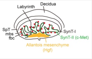
It turns out that the migration depends on hepatocyte growth factor (HGF) and c-Met signaling axis.
This work first established the in vitro SynT-II cell differentiation protocol, which is helpful to dissect how the placental interface established.
Furthermore, it suggests that the migration of SynT-II cells might contribute to the formation of the elaborate structure of labyrinth, under the regulation of HGF/c-Met signaling.
This work is supported by grants from National Natural Science Foundation of China and the Ministry of Science and Technology of China.
A diagram illustrating a mature mouse placenta and expression patterns of c-Met (green) and Hgf (yellow). (Image by IGDB)