Microglia are the primary immune cells in the brain. Under pathological conditions, they become activated and participate in scavenging, inflammation and tissue repair in response to brain injury. The functions and underlying mechanisms of activated microglia have been intensively studied and well understood. Under physiological conditions, microglia typically stay in a "resting" state during most of the time, with highly branched processes continuously extending to and retracting from surrounding brain tissues on a time scale of minutes. Whether and how such highly dynamic resting microglia functionally interact with neurons remain to be elucidated.
A team led by Dr. DU Jiulin at the Institute of Neuroscience and State Key Laboratory of Neuroscience, Shanghai Institutes for Biological Sciences identified the physiological function of resting microglia—a major breakthrough in understanding the role of microglia in the healthy brain.
In this study, by combining in vivo time-lapse confocal and two-photon imaging, glutamate uncaging, whole-cell recording and FRET imaging, researchers simultaneously monitored, for the first time, both the motility of resting microglialprocesses and the activity of surrounding neurons in intact zebrafish optic tectum, and examined the interaction between them.
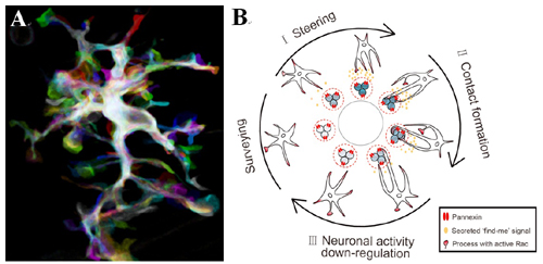
They found that locally elevated neuronal activity steers resting microglial processes towards the soma of highly active neurons and facilitates the formation of microglia-neuron contact. This process requires the activation of pannexin-1 hemichannels in neurons and of small Rho GTPase Rac in resting microglia. Interestingly, they found that such microglia-neuron contact in turn down-regulates both spontaneous and visually evoked activities in the contacted neuron.
This study reveals an instructive role of neuronal activity in resting microglial motility and suggests a novel function of microglia in homeostatic regulation of neuronal activity in the healthy brain.
This work entitled "Reciprocal Regulation between Resting Microglial Dynamics and Neuronal ActivityIn Vivo" was published online in Developmental Cell on Nov. 29, 2012.
The research was supported by grants from the Ministry of Science and Technology of China, Chinese Academy of Sciences, and Shanghai Municipal Government.
CONTACT:
DU Jiulin
Phone: 86-21-54921825
E-mail: forestdu@ion.ac.cn
Institute of Neuroscience and State Key Laboratory of Neuroscience,
Shanghai Institutes for Biological Sciences, Chinese Academy of Sciences,
Shanghai, China

A. Overlay of projected images of a resting microglial cell acquired every 5 min for an hour during in vivo time-lapse confocal imaging of the optic tectum in a 6-dpf Tg (Apo-E:eGFP) zebrafish larva. B. Working model for the bidirectional modulation between resting microglia and neuron. (Image by Dr. DU Jiulin's group)

86-10-68597521 (day)
86-10-68597289 (night)

86-10-68511095 (day)
86-10-68512458 (night)

cas_en@cas.cn

52 Sanlihe Rd., Xicheng District,
Beijing, China (100864)

