
In romantic literature, every relationship – even true love – is said to comprise love and hate without exception. This law also goes for nature, as evidenced by the “love-hate relationship” between plants and sunlight. On the one hand, plants absolutely must receive sunlight in order to undertake photosynthesis. On the other hand, excessive sunlight causes irreversible damage to the photosynthetic machinery of plants.
To reduce or avoid such photooxidative damage, plants have evolved an efficient photoprotective mechanism called energy-dependent quenching (qE). Under high light conditions, the lumenal pH of plant thylakoid drops from 6.5 to 5.5–5.8, leading to the activation of PsbS, a photosystem II protein embedded in the thylakoid membrane. The activated PsbS then initiates qE, enabling plants to safely dissipate excess light energy as heat.
The essential role of PsbS in qE was reported in 2000. However, its pigment-binding properties are still controversial, and its mechanism of action remains elusive. Therefore, solving the three-dimensional structure of PsbS has been long awaited in the photosynthesis field. Four years ago, CHANG Wenrui’s group at the Institute of Biophysics, Chinese Academy of Sciences, embarked on the structural investigation of PsbS to elucidate the mechanism of PsbS action in qE.
In a new study published online August 10 in Nature Structural & Molecular Biology, entitled Crystal Structures of the PsbS Protein Essential for Photoprotection in Plants, CHANG’s group released their structural and functional analysis of plant photoprotective protein PsbS.
As part of their study, the researchers developed a protocol by which a large quantity of homogeneous PsbS protein could be purified from spinach leaves. They purified and crystallized PsbS at low pH and solved its structure in the active state at 2.35 Å resolution. The crystal structure reveals that PsbS is composed of four transmembrane helices that form a compact structure, thus leaving no internal space for formation of pigment-binding sites within the PsbS monomer.
The overall structure of PsbS is significantly different from the structure of LHCII and CP29, two members of the LHC superfamily whose structures had previously been solved by CHANG’s group. Combining structural analyses and biochemical experiments, they demonstrated that PsbS is a compact dimer at low pH (in the active state).
Moreover, based on the structural analysis of qE inhibitor DCCD-bound PsbS, together with biochemical experiments, they found that PsbS is a dimer in both inactive and active states, and the transition between the two states involves low pH-induced conformational change of the lumenal loops of the PsbS dimer. Interestingly, they also found a chlorophyll a molecule at the PsbS dimer interface, which indicates that PsbS may participate in qE directly.
Their findings provide important insights into the mechanism of PsbS activation and inhibition and a putative mechanism of PsbS action in qE. The reviewers of the manuscript spoke highly of the research and made the following comments: “The present work clearly represents an essential step forward”; “it represents a breakthrough in the understanding of the structural basis of photo protection in plants”; and “obtaining a structure of PsbS is a major breakthrough.”
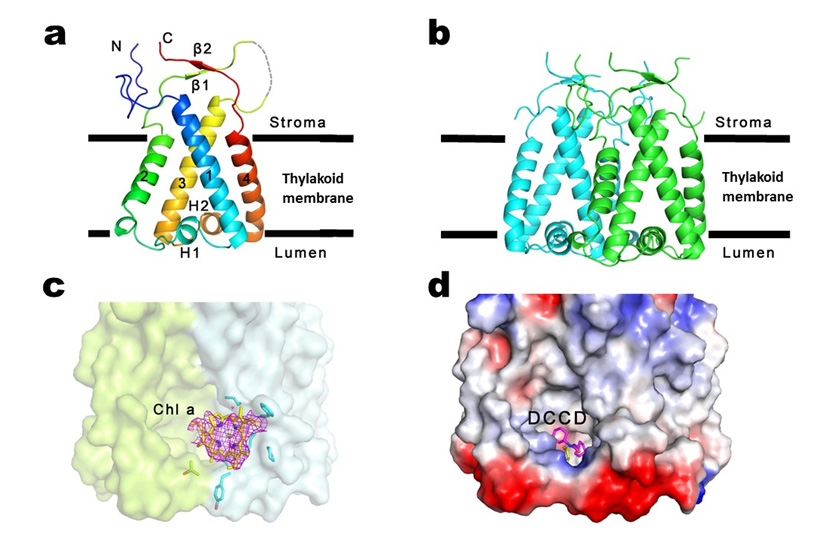
Overall structures of PsbS: a. PsbS monomer; b. PsbS dimer; c. the potential chlorophyll a molecule at the PsbS dimer interface; d. the qE inhibitor DCCD bound to the PsbS dimer (Image by CHANG Wenrui’s research group)

86-10-68597521 (day)
86-10-68597289 (night)

52 Sanlihe Rd., Xicheng District,
Beijing, China (100864)
Copyright © 2002 - Chinese Academy of Sciences


