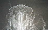
A phase contrast image of a fly head obtained by CAS researchers.
A research team from the CAS Institute of High Energy Physics (IHEP) has made progress with its work on the phase contrast imaging by using hard X-rays.
Conventional x-ray imaging techniques, which have been in operation for over 100 years, utilize differences in sample absorption to yield image contrast. A new area of X-ray science, called phase contrast imaging, can provide great enhancements in the image. In the hard X-ray (10-100 keV) regime, phase-enhanced techniques can improve the image contrast by orders of magnitude.
After repeated experiments, CAS researchers at the BL 4W1A Topography Station of the Beijing Synchrotron Radiation, IHEP, succeeded in obtaining nice phase contrast images of different specimens by using hard X-rays through the diffraction enhancement method.
The development of this new technology has the potential to provide new applications in medical, industrial and biological environments, says researchers. It could revolutionize current imaging and inspection systems.


