Newsroom


A new study published in PNAS on August 21 reveals that a distinct population of macrophages can directly "eat" scar-forming material in the liver and promote the reversal of liver fibrosis. The finding reveals an unrecognized mechanism by which macrophages help clear fibrotic tissue: not by secreting factors, but by physically removing the extracellular matrix (ECM).
The study was jointly conducted by researchers from the Institute of Biophysics of the Chinese Academy of Sciences, the First Affiliated Hospital of Sun Yat-sen University, and the First Affiliated Hospital of Guilin Medical University.
Liver fibrosis is a common pathological pathway underlying most chronic liver diseases. At its core, it is characterized by the excessive accumulation of extracellular matrix (ECM), which forms scar tissue in the liver. Macrophages play a central regulatory role in both the development and regression of liver fibrosis. However, the question of how macrophages switch between promoting scar formation and driving scar resolution has long remained unanswered.
The researchers identified a distinct macrophage subset, termed ReM2, with a unique phagocytic function that enables it to directly "eat" scar tissue in the liver.
Through an integrated analysis of single-cell RNA sequencing and spatial transcriptomics, the researchers discovered that ReM2 cells emerged during liver fibrosis progression and continued to increase in number during fibrosis regression. Eventually, they became the most abundant subset of macrophages in the liver during the regression phase, exceeding the number of profibrotic scar-associated macrophages (SAMs) by more than tenfold.
Spatial mapping further revealed that ReM2 cells specifically accumulated in fibrotic microenvironments enriched in collagen deposition, suggesting direct physical interactions between ReM2 cells and scar tissue.
The study showed that ReM2 clears ECM through two receptor-mediated pathways. First, ITGA4 directly recognizes and binds to fibronectin. Second, FCGR4 recognizes collagen I tagged by endogenous IgG2a antibodies, thereby mediating antibody-dependent phagocytic clearance.
Immunoprecipitation experiments, in vivo functional studies, and genetic approaches provided compelling evidence that FCGR4 and ITGA4 are indispensable for the scar-clearing activity of ReM2.
Based on these findings, the researchers proposed a new model of receptor-mediated phagocytic clearance of ECM by macrophages. In this model, ReM2 macrophages directly recognize and engulf collagen I and fibronectin through surface receptors FCGR4 and ITGA4, thereby actively driving the reversal of liver fibrosis.
This study precisely defined the molecular characteristics, spatiotemporal dynamics, and functional mechanisms of ReM2 at single-cell and spatial resolution. These findings provide a new conceptual framework for understanding how tissue fibrosis can be actively reversed and identify potential therapeutic targets, including FCGR4 and ITGA4, for the development of anti-fibrotic therapies.
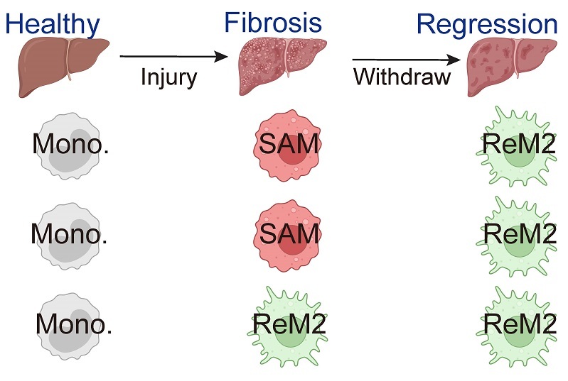
Schematic model of the SAM/ReM2 macrophage populations. Under homeostatic conditions, monocytes are the predominant population. Upon injury, monocytes differentiate primarily into SAMs, accompanied by a small proportion that differentiates toward ReM2. During the recovery phase, SAMs rapidly decline, while ReM2 cells continue to accumulate. (Image by ZHOU Haining's group)
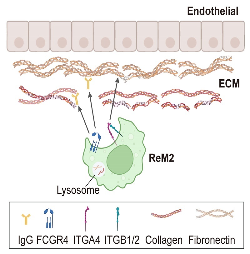
Mechanistic model of ReM2-mediated ECM degradation. ReM2 mediates ECM degradation through ITGA4-FN binding and FCGR4-mediated recognition of IgG2a-tagged FN/collagen I complexes. (Image by ZHOU Haining's group)