
Newsroom


Autophagy is the process by which cells remove damaged proteins, recycle worn-out organelles (e.g., mitochondria), clear cellular waste, and provide nutrients during stress. Autophagy is essential for muscles because they are constantly under mechanical stress. If autophagy is too low, damaged proteins accumulate and muscle gradually weakens. If it is too high, muscle tissue can begin breaking itself down.
Disruption of autophagy has been implicated in a wide range of muscle disorders, and abnormal muscle autophagy is frequently observed in neurogenic diseases. However, the neuronal signaling pathways that control this process had previously remained largely unknown.
Now, researchers led by Prof. ZHANG Hong from the Institute of Biophysics of the Chinese Academy of Sciences have identified two parallel neuronal circuits that regulate the autophagy-lysosome pathway in the body wall muscle of Caenorhabditis elegans, a tiny nematode worm. Their research has uncovered a previously unknown mechanism by which the worm’s nervous system maintains muscle homeostasis.
The findings, published in Developmental Cell on June 29, provide new insights into neuron-to-muscle communication and may inform future therapies for neurogenic muscle disorders such as amyotrophic lateral sclerosis (ALS) and spinal muscular atrophy (SMA).
In this study, the researchers found that the first neuronal circuit is formed by electrical synapses between AVA interneurons and A-type motor neurons (A-MNs), which use the proteins UNC-7/UNC-9 to form synaptic gap junctions. Loss of these electrical synapses promotes the release of the neuropeptides NLP-9, NLP-12, and FLP-18. Among them, NLP-12 and FLP-18 activate the NPR-5/Gαq–EGL-30 signaling pathway in body wall muscle, increasing intracellular Ca2+ levels.
The second neuronal circuit originates from ASI sensory neurons, where the TGF-β-like signaling molecule DAF-7 activates the canonical TGF-β signaling pathway in the body wall muscle to regulate intracellular Ca2+ homeostasis.
Together, these two parallel neuronal pathways converge on a Ca2+–calpain–lysosome signaling cascade to maintain autophagic activity in the body wall muscle of C. elegans, thereby preserving muscle structure and function.
The researchers also found that unc-7 and daf-7 mutants exhibited severe disruption of muscle fiber organization and significantly reduced swimming ability, both characteristic features of myopathy. Genetic restoration of the affected signaling pathways markedly alleviated autophagy defects and restored muscle structural integrity and function.
The study uncovers a previously unrecognized mechanism of neuron-to-muscle communication and offers new insights into the pathogenesis of neurogenic myopathies, potentially opening new avenues for therapeutic development.
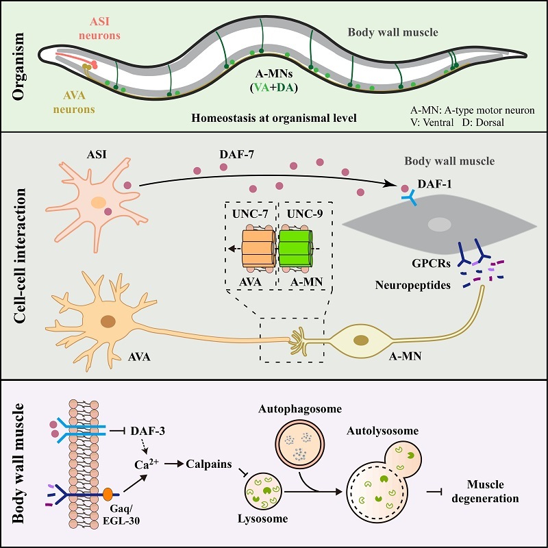
Model for how neuron-to-muscle singling orchestrates muscle autophagy and homeostasis (Image by ZHANG Hong's group)
